

articles
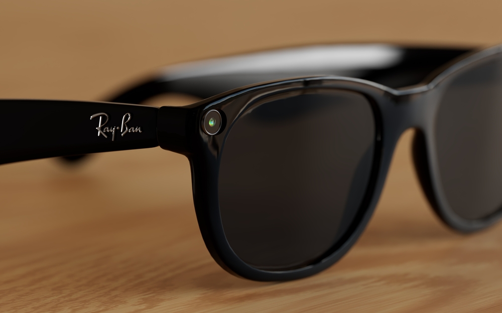
Ray-Ban has long been recognized as a leader in timeless style, but now the brand is taking a bold step into the future. In collaboration with Meta, Ray-Ban has introduced a groundbreaking innovation - Ray-Ban Meta Glasses. These glasses are a fusion of iconic design and next-generation technology that allows you to capture, share, and stay connected like never before.
What Are Ray-Ban Meta Glasses?
Ray-Ban Meta Glasses go beyond vision correction or sun protection - they seamlessly integrate a camera, speakers, and voice-activated AI assistance, allowing users to capture, share, and interact hands-free. With their classic Ray-Ban frames, these glasses look just like traditional eyewear, but they’re packed with cutting-edge features hidden in plain sight.
Key Features and Technology
Built-in Cameras: Capture photos and videos instantly with a simple voice command or tap on the frame. The dual 12MP cameras allow for high-quality media with depth and detail.
Open-Ear Audio: Discreet speakers positioned near your ears provide clear sound without blocking ambient noise, letting you listen to music, take calls, or hear notifications seamlessly.
Meta AI Integration: Access Meta’s AI assistant through voice commands. Ask questions, control your device, or get updates - all without taking your phone out.
Live Streaming and Sharing: Stream directly to social media platforms like Instagram and Facebook in real time, making it easy to share your perspective with your audience.
Touch and Voice Controls: Intuitive touch-sensitive temples and responsive voice commands give you full control over your smart features.
Transitions® Lenses Option: Enjoy lenses that automatically adapt to changing light conditions for optimal comfort both indoors and outdoors.

Our eyes are extremely delicate, yet they can be subjected to harsh conditions and other environmental factors that affect their health. One of the problems that can affect our eyes is an accumulation of dirt, debris and bacteria on the eyelids. This can cause a range of issues, including stopping tear film from reaching the eyes and being properly dispersed over their surface – which is necessary to keep them healthy and comfortable. Fortunately, a new solution called Blephex® can help.
What is Blephex®?
Blephex® is a handheld electro-mechanical device that is applied to the margins of the eyelids with the purpose of cleaning them and improving the effectiveness with which tear film flows onto the surface of the eyes.
Blephex® has a disposable, surgical-grade sponge tip which rapidly oscillates to create a cleaning action. Before the sponge tip is placed onto the eyes, it is soaked in a gentle exfoliating solution. This solution provides soft abrasion to help remove dead skin cells and debris that could be irritating the eyes and interrupting tear film progression. The Blephex® device is manually applied to the eyes and moved gently across the eyelids, with the entire, painless process taking approximately 6 to 8 minutes per eye. A different sponge is used on each eye, ensuring that no bacteria is passed between them. After the procedure, patients are given instructions on how to maintain the cleanliness of their eyelids with daily/nightly eyelid hygiene at home.
Most patients experience a significant improvement in tear film production and dispersal, and a reduction in unpleasant symptoms that they may have been experiencing within 48 hours of their treatment. While a single treatment is normally enough to produce excellent results, many patients are advised to have Blephex® every 4-6 months.

In September 2025, the FDA approved Stellest lenses for children ages 6 to 12 to correct and manage myopia, a major milestone for myopia care. These lenses have been successfully used in countries across Europe, Canada, and Asia since 2020, showing promising results in slowing eye growth associated with myopia.
The Importance of Early Myopia Management
Myopia typically begins in early childhood, often between the ages of 6 and 10, and tends to worsen quickly during the school years as children spend more time reading and using digital devices. The sooner it begins, the higher the risk that it will progress to more severe levels. Every increase in myopia level significantly raises the risk of eye health complications later in life, such as retinal detachment or glaucoma.
By beginning myopia management as soon as possible, parents can help slow eye growth and protect long-term vision. With innovative options like Stellest lenses, families now have effective, science-backed solutions to take control of their child’s myopia.
What Are Stellest Lenses?
Stellest lenses are a groundbreaking innovation in myopia control, developed by Essilor. They look like regular eyeglass lenses but are designed with specialized optical technology to both correct vision and slow the progression of myopia.

Dry eye is a common condition that affects millions of individuals worldwide. It occurs when the eyes do not produce enough tears or the tears evaporate too quickly. This can lead to discomfort, irritation, and even vision problems. TearCare offers a breakthrough treatment option for long-lasting relief from dry eye.
What is TearCare?
TearCare is a revolutionary treatment option that offers long-lasting relief for dry eye sufferers. This innovative technology utilizes wearable eyelid warming devices to precisely deliver heat to the meibomian glands, effectively unclogging them and promoting healthier oil production. The treatment is performed in-office by a trained eye care professional and typically takes less than 15 minutes.
TearCare targets the root cause of the problem - meibomian gland dysfunction. By restoring the proper function of these glands, TearCare helps to stabilize the tear film, improve tear production, and alleviate the symptoms of dry eye. This breakthrough solution offers a safe and effective alternative to medications and artificial tear drops, providing long-lasting relief for those suffering from dry eye.
How TearCare Works to Treat Dry Eye
TearCare works by gently applying heat to the eyelids, stimulating the meibomian glands and promoting the release of healthy oils. The wearable eyelid warming devices are designed to maintain a consistent temperature, ensuring optimal results. The heat helps to liquefy any hardened oils or debris clogging the glands, allowing them to function properly again. This treatment can also help reduce inflammation and improve overall eyelid hygiene.

If you have recently undergone eye surgery, your eye doctor may recommend the use of post-surgical contact lenses as part of your recovery process. These specialized lenses are designed to aid in the healing and protection of your eyes after surgery.
Why Post-Surgical Contact Lenses May Be Necessary
After eye surgery, your eyes are in a delicate state and require extra care to ensure proper healing. Post-surgical contact lenses can play a crucial role in this process. These lenses act as a protective barrier, shielding your eyes from irritants such as dust, debris, and bright lights. They also help to maintain the shape of the cornea and aid in the prevention of infection. By wearing post-surgical contact lenses, you can minimize the risk of complications and promote a faster and smoother recovery.
Different Types of Post-Surgical Contact Lenses
There are various types of post-surgical contact lenses available, and the specific type recommended for you will depend on the nature of your surgery and your eye condition. One common type is the bandage contact lens, which is a soft, therapeutic lens that covers the cornea, providing protection and promoting healing.
Another type is scleral lenses, which is larger and covers a larger portion of the eye, providing enhanced protection and support. Your optometrist will determine the most suitable type of post-surgical contact lens for your individual needs.

Visual field testing is an important part of most standard comprehensive eye exams. Also sometimes known as perimetry testing, Visual field testing is a method to measure the entire scope of vision of an individual, including their peripheral/side vision.
The importance of visual field testing
Visual field testing is one of the most effective diagnostic treatments in the detection of glaucoma. This is because when patients are affected by glaucoma, it is usually the peripheral vision that is affected by their condition first. However, it can also be used to detect central or peripheral retinal diseases, eyelid conditions such as drooping, optic nerve damage and conditions that affect the visual pathways from the optic nerve to the area of the brain where this information is processed into vision.
Visual field testing is also an important part of monitoring for people who are considered to be at risk for vision loss from disease and other problems, including those who have been diagnosed with the following:
Multiple sclerosis
Hyperthyroidism
Pituitary gland disorders
Central nervous system problems (such as a tumor that may be pressing on the brain)
Stroke
Diabetes
High blood pressure
Neuro-Optometric Rehabilitation
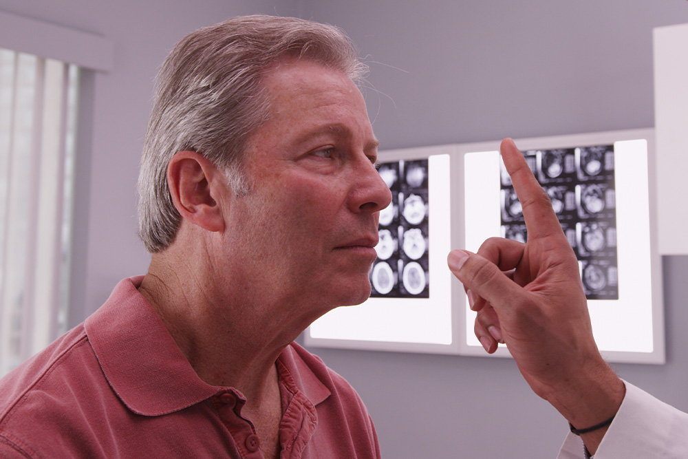
When most people think about vision problems, they picture issues with the eyes themselves. However, many visual difficulties actually begin in the brain. After a concussion, stroke, or other brain injury, the connection between the eyes and brain can be disrupted - causing symptoms that affect balance, reading, focus, and even daily comfort.
Brain Injuries and Vision
Your visual system is complex - about 70% of the brain is involved in vision processing. After a traumatic brain injury (TBI), concussion, or neurological event such as a stroke, the pathways that coordinate eye movement, focus, and perception can become impaired.
This can result in symptoms such as:
Blurred or double vision
Eye strain and headaches
Difficulty reading or focusing
Dizziness or balance problems
Light sensitivity
Problems with depth perception
These visual issues often persist long after other symptoms of a brain injury have improved, making specialized vision care essential for recovery.
What Is Neuro-Optometric Rehabilitation?
Neuro-optometric rehabilitation is a specialized form of vision therapy designed to retrain how the brain and eyes work together. Unlike traditional eye care that focuses on eyesight alone, this therapy targets the neurological processes behind vision. Our doctor evaluates how the eyes and brain communicate and creates a personalized treatment plan to rebuild these visual pathways. The goal is to restore comfort, coordination, and clarity - improving overall visual function and quality of life.

Dry eye is a common condition that occurs when your eyes do not produce enough tears or when the tears evaporate too quickly. This can result in discomfort, irritation, and a gritty sensation in the eyes. Understanding the causes and symptoms of dry eye is crucial for finding effective treatment options. Tyrvaya offers a breakthrough solution for dry eye relief.
What Causes Dry Eye?
There are several factors that can contribute to the development of dry eye. The meibomian glands are responsible for producing the oily component of the tear film, which helps prevent evaporation of tears and maintains a smooth ocular surface. Meibomian gland dysfunction occurs when these glands become blocked, leading to a decrease in the quantity and quality of the meibum. This can result in evaporative dry eye, discomfort, and inflammation of the eyelid margins.
Another common causes is age. As we get older, our tear production tends to decrease, making us more prone to dryness. Hormonal changes, such as those that occur during menopause, can also affect tear production and lead to dry eye.
Environmental factors can play a role as well. Dry or windy climates, air conditioning, and excessive screen time can all contribute to dry eye. Additionally, certain medications, such as antihistamines and antidepressants, can cause dryness as a side effect.
Other underlying health conditions, such as autoimmune diseases like Sjogren's syndrome or rheumatoid arthritis, can also contribute to dry eye. In these cases, the body's immune system mistakenly attacks the tear glands, leading to reduced tear production.

Migraines are a debilitating neurological condition that affect millions of people worldwide. One of the most common and disabling symptoms of migraines is light sensitivity, also known as photophobia. This heightened sensitivity to light can be a significant source of discomfort and pain for those suffering from migraines, making it difficult to function during an attack.
Photophobia is a common symptom of migraines, with up to 80% of migraine sufferers experiencing sensitivity to light during an attack. This sensitivity can be triggered by various types of light, including natural sunlight, fluorescent lighting, and even the glow of computer screens or smartphones.

In the realm of eye care, surgical co-management has emerged as a collaborative approach that aims to provide patients with comprehensive and seamless treatment. This concept involves the joint efforts of optometrists and ophthalmologists, each bringing their unique expertise to the table. By working together, these eye care professionals strive to enhance patient outcomes and deliver exceptional care.
The Role of Optometrist and Ophthalmologist in Surgical Co-Management
Surgical co-management is built upon the unique skill sets and areas of expertise of optometrists and ophthalmologists. By understanding their respective roles, you can appreciate the synergy that this collaborative approach fosters.
Optometrists are primary eye care professionals who specialize in the examination, diagnosis, and non-surgical treatment of vision disorders. Their responsibilities in surgical co-management include:
Performing comprehensive eye examinations and evaluations
Monitoring and managing pre-existing eye conditions
Providing pre-operative and post-operative care
Educating patients on surgical procedures and aftercare
Collaborating with ophthalmologists to ensure continuity of care
Ophthalmologists are medical doctors who specialize in the diagnosis, treatment, and surgical management of eye diseases and disorders. Their role in surgical co-management encompasses:
Evaluating patients' candidacy for surgical interventions
Performing complex surgical procedures
Providing specialized medical and surgical care
Collaborating with optometrists to ensure seamless patient care
Monitoring and managing post-operative complications
By combining the expertise of optometrists and ophthalmologists, surgical co-management ensures that patients receive comprehensive and coordinated care throughout their treatment journey.
How Surgical Co-Management Works
Surgical co-management is a well-orchestrated process that involves several key steps. Understanding how it works can help you navigate this collaborative approach with confidence.
Initial Evaluation: The process typically begins with an optometrist conducting a comprehensive eye examination. During this evaluation, the optometrist assesses the patient's visual needs, identifies any potential issues, and determines if a surgical intervention is necessary.
Referral and Consultation: If surgery is recommended, the optometrist refers the patient to an ophthalmologist for further evaluation and consultation. This step ensures that the patient receives specialized medical advice and a thorough assessment of their suitability for the proposed surgical procedure.
Pre-operative Care: The optometrist plays a crucial role in providing pre-operative care, which may include managing any existing eye conditions, ensuring the patient understands the surgical process, and addressing any concerns or questions they may have.
Surgical Procedure: The ophthalmologist performs the necessary surgical intervention, leveraging their specialized training and expertise in surgical techniques.
Post-operative Care: After the surgery, the patient's care transitions back to the optometrist, who closely monitors the recovery process and provides post-operative care and management. This may involve follow-up appointments, monitoring for any complications, and ensuring the patient adheres to the prescribed treatment plan.
Ongoing Collaboration: Throughout the entire process, the optometrist and ophthalmologist maintain open communication and collaborate closely. This ensures that the patient's care is seamless, and any concerns or issues are promptly addressed by the appropriate healthcare professional.


Contact Info
Office Address
Fax Number
(760) 727-3044
(760) 727-3044
Office Hours
- Monday 9:00am - 6:00pm
- Tuesday 9:00am - 6:00pm
- Wednesday 7:00am - 5:00pm
- Thursday 9:00am - 6:00pm
- Friday 9:00am - 2:00pm
- Saturday Closed
- Sunday Closed
© 2026 Shadowridge Family Vision Center. All rights Reserved. Accessibility Statement - Privacy Policy - Sitemap
Powered by:
