

articles
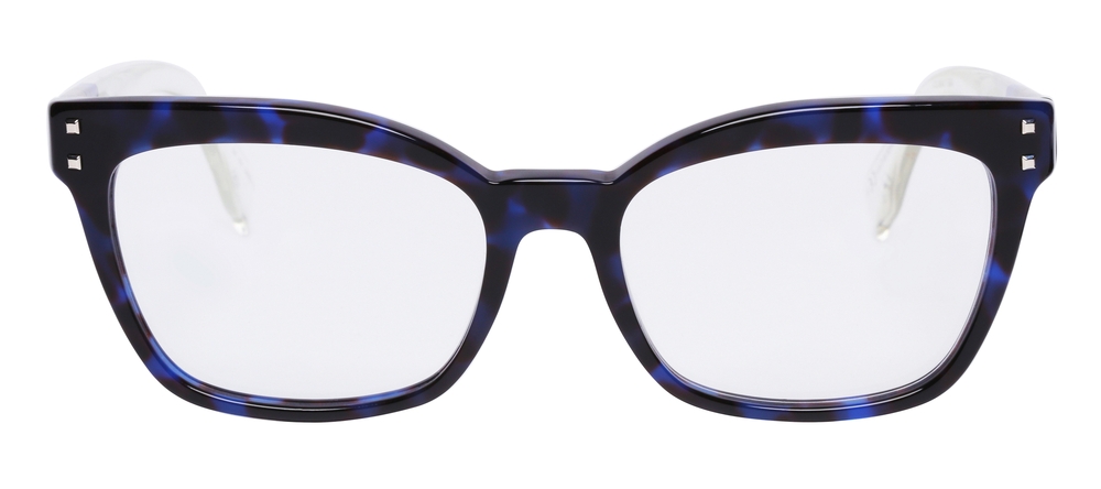
Technology continues to blur the lines between style and functionality - and Nuance Audio Glasses are a perfect example of this evolution. Designed to look like everyday eyewear, these innovative glasses feature built-in hearing enhancement technology that helps users hear conversations clearly while maintaining natural, effortless vision.
What Are Nuance Audio Glasses?
Nuance Audio Glasses are smart eyeglasses designed to provide both vision correction and enhanced hearing. Developed by Nuance Hearing, these glasses discreetly integrate directional microphones, miniature speakers, and Bluetooth connectivity into the frame - allowing wearers to improve their hearing experience without traditional hearing aids. By combining hearing technology with fashionable eyewear, Nuance Audio Glasses offer a seamless, everyday solution for individuals who want better hearing support without drawing attention to their device.
How Nuance Audio Glasses Work
The technology behind Nuance Audio Glasses focuses on selective sound amplification and speech clarity. The glasses use beamforming microphones to focus on voices in front of the wearer while reducing background noise. The enhanced sound is then transmitted through tiny speakers near the ears, allowing users to hear clearly without the occlusion effect common with in-ear devices.
This open-ear design keeps the ear canals unobstructed, promoting natural hearing while boosting important speech frequencies - ideal for conversations in noisy environments like restaurants or social gatherings.

Senior eye care is crucial for maintaining optimal vision and overall eye health as we age. As individuals grow older, they become more susceptible to age-related eye conditions. Regular comprehensive eye exams play a key role in early detection and management of these conditions, helping to preserve vision and prevent potential vision loss. By staying proactive with eye care and seeking timely professional assistance, seniors can enjoy clear, comfortable vision and ongoing eye health for years to come.
Common Eye Conditions in Seniors
As we reach our golden years, there are several eye conditions that become more prevalent. These conditions can have a significant impact on our vision and overall quality of life.
Presbyopia: This condition affects near vision, making it difficult to focus on close objects. It is a normal part of aging and is typically addressed with reading glasses or multifocal lenses.
Floaters and Flashes: As the vitreous inside the eye changes with age, it can lead to the perception of floaters (small dark spots or lines) and flashes of light. While often harmless, sudden onset of floaters and flashes may indicate a retinal tear or detachment and should be promptly evaluated.
Age-Related Macular Degeneration (AMD): AMD is a leading cause of vision loss in older adults. It affects the macula, the central part of the retina, leading to blurred or distorted central vision. There are two forms of AMD, dry AMD, and wet AMD, with the latter being more severe.
Cataracts: Cataracts are a common age-related condition characterized by the clouding of the eye's natural lens. This can lead to blurry vision, glare, and difficulty seeing in low-light conditions.
Glaucoma: Glaucoma is a group of eye conditions that damage the optic nerve, often due to elevated intraocular pressure. It can result in peripheral vision loss and, if left untreated, may lead to total blindness.
Diabetic Retinopathy: For seniors with diabetes, diabetic retinopathy is a concern. It occurs when high blood sugar levels damage the blood vessels in the retina, leading to vision problems and potential blindness if not managed.
Dry Eye Syndrome: Seniors may experience dry eye syndrome, characterized by insufficient lubrication and moisture on the surface of the eye. It can cause discomfort, redness, and fluctuating vision.

If you find it difficult to tell colors apart, you may be color blind. Color blindness, or color deficiency, is estimated to affect around 8% of men and about 1% of women, but for those affected, it can significantly impact the quality of their day-to-day life. Contrary to popular belief, being color blind doesn’t mean that you can’t see any color at all. Instead, patients simply struggle to differentiate between certain colors. The vast majority of people who are color blind find it impossible to tell the difference between varying shades of red and green. You may hear this referred to as red-green color deficiency. However, this doesn’t only mean that they mix up red and green. They can also mix up colors that have some green or red light as part of their whole colors, for example purple and blue. This is because they are unable to see the red light that forms part of the color purple.
As you can probably imagine, this type of visual impairment can be a problem for things like traffic lights, taking medications and even looking at signs and directions. For example, someone who is color blind may find that the green on a traffic light may appear white or even blue.

Strabismus is a relatively common condition, affecting about 2-4% of the population worldwide. It is one of the most frequent eye disorders seen in children, though it can also develop in adults due to conditions like stroke or trauma. In the United States alone, it is estimated that around 12 million people are affected by strabismus, with varying degrees of severity. Early diagnosis and treatment are essential, as untreated strabismus in children can significantly impair visual development.
What is Strabismus?
Strabismus, commonly referred to as crossed eyes, is a condition in which the eyes do not properly align with each other when looking at an object. One eye may turn in, out, up, or down, while the other eye focuses correctly. This misalignment can be constant or intermittent and may affect one or both eyes. Strabismus occurs when the muscles controlling eye movement do not work together correctly, leading to difficulty in focusing both eyes on a single point. This condition can lead to visual issues such as double vision and, in some cases, can result in amblyopia (lazy eye) if left untreated.
How Does Strabismus Develop?
Strabismus develops when there is a disruption in the coordination of the muscles that control eye movement, leading to a misalignment of the eyes. This disruption can result from various causes, including genetic factors, neuromuscular conditions, or problems with the nerves that transmit signals to the eye muscles. In some cases, it may be present at birth, while in others, it can develop later in childhood or adulthood due to conditions such as cerebral palsy, Down syndrome, or even trauma to the eye or brain.
Signs and Symptoms of Strabismus
The primary symptom of strabismus is the visible misalignment of the eyes. However, there are other signs and symptoms that may indicate the presence of this condition:
Blurred or double vision
Headaches or eye strain
Difficulty with depth perception and spatial awareness
Reduced visual acuity in one eye
Frequent eye rubbing or squinting
Tilting or turning the head to compensate for the misalignment

As technology continues to advance, so does the field of the optometric industry. The development of innovative tools and techniques has allowed for more accurate and comprehensive examinations. One such technology is Optos, a revolutionary system that utilizes ultra-widefield retinal imaging technology to provide optometrists with a detailed view of the entire retina.
How Does Optos Work?
Optos technology is based on the principle of ultra-widefield retinal imaging, which allows for a wider and more detailed view of the retina compared to traditional imaging techniques. The Optos system consists of a specialized camera that captures high-resolution images of the retina using scanning laser ophthalmoscopy (SLO) and optical coherence tomography (OCT) technologies. SLO provides a wide-field view of the retina, while OCT allows for cross-sectional imaging, providing valuable insights into the various layers of the retina.
The Optos camera is designed to capture images through a process called optomap, which captures up to 200 degrees of the retina in a single image. This wide-field view provides optometrists with a comprehensive picture of the retina, enabling them to detect abnormalities that may not be visible with traditional imaging techniques. The optomap image can be instantly viewed and analyzed by your eye doctor, allowing for a more efficient and accurate diagnosis.
Common Eye Conditions Detected by Optos
Optos technology has the capability to detect a wide range of eye conditions, including but not limited to, diabetic retinopathy, macular degeneration, glaucoma, and retinal tears or detachments. Diabetic retinopathy is a condition that affects individuals with diabetes, causing damage to the blood vessels in the retina. Optos can capture detailed images of the retina, enabling optometrists to detect any signs of diabetic retinopathy and initiate appropriate treatment.
Macular degeneration is another common eye condition that can be detected using Optos. This condition affects the macula, the central part of the retina responsible for sharp, central vision. Optos allows for a comprehensive view of the macula, identifying any changes or abnormalities that may indicate the presence of macular degeneration.
Glaucoma, a condition characterized by damage to the optic nerve, can also be detected using Optos. The wide-field view provided by Optos allows for a thorough examination of the optic nerve and the surrounding structures, facilitating early detection and intervention.
Finally, Optos technology is particularly effective in detecting retinal tears or detachments. These conditions can lead to sudden vision loss and require immediate medical attention. Optos allows for a comprehensive view of the retina, identifying any signs of retinal tears or detachments and initiate prompt treatment.

Astigmatism is a common refractive error that occurs when the cornea or lens of the eye is not perfectly spherical. Instead of having a uniform, rounded shape, the cornea or lens has a slightly more curved or irregular shape, resembling more of an oval or football-like appearance.
This irregular shape causes light to bend or refract unevenly as it enters the eye, leading to blurred or distorted vision. Astigmatism is often present from birth, but it can also develop later in life due to various factors, such as eye injuries, certain medical conditions, or the natural aging process.
Common Symptoms of Astigmatism
If you have astigmatism, you may experience a range of symptoms that can impact your daily life. Some of the most common symptoms of astigmatism include:
Blurred or distorted vision, especially at a distance or when reading
Difficulty seeing clearly at night or in low-light conditions
Frequent headaches or eye strain
Sensitivity to glare or bright lights
Double vision or the appearance of halos around lights
These symptoms can vary in severity and may become more pronounced over time, especially if the astigmatism is left uncorrected.

Post-concussion syndrome (PCS) is a complex condition that can occur after a traumatic brain injury (TBI), such as a concussion. While the initial injury may have healed, the lingering symptoms can significantly impact your daily life, making it challenging to return to your normal routines and activities.
What is Post-Concussion Syndrome?
Post-concussion syndrome is a collection of physical, cognitive, and emotional symptoms that can persist for weeks, months, or even years after a head injury. Unlike the typical recovery from a concussion, which usually resolves within a few days or weeks, PCS can be a prolonged and debilitating experience. The symptoms of PCS can vary widely from person to person, making it a complex and often misunderstood condition.
Causes of Post-Concussion Syndrome
One of the primary causes of post-concussion syndrome is the disruption of the brain’s normal function due to the injury itself. When the brain experiences trauma, it can lead to a variety of biochemical and structural changes, which may cause the ongoing symptoms of PCS. The severity of the concussion does not always correlate with the likelihood of developing PCS; even minor concussions can lead to prolonged post-concussion symptoms in some individuals.
Another contributing factor to PCS may be related to the individual’s psychological response to the injury. Anxiety, stress, and pre-existing mental health conditions can exacerbate the symptoms of PCS and prolong the recovery process. People who have a history of migraines, anxiety disorders, or depression may be more prone to developing post-concussion syndrome.
Recognizing the Symptoms of Post-Concussion Syndrome
The symptoms of post-concussion syndrome can be diverse and can vary in severity from person to person. Some of the most common symptoms include:
Physical Symptoms: Headaches, dizziness, nausea, sensitivity to light or sound, fatigue, and sleep disturbances.
Cognitive Symptoms: Difficulty concentrating, memory problems, slower processing speed, and reduced problem-solving abilities.
Emotional Symptoms: Irritability, mood swings, anxiety, depression, and increased emotional sensitivity.
The presence and severity of these symptoms can fluctuate over time, making it crucial to monitor your condition closely and communicate any changes to your primary care or eye doctor.
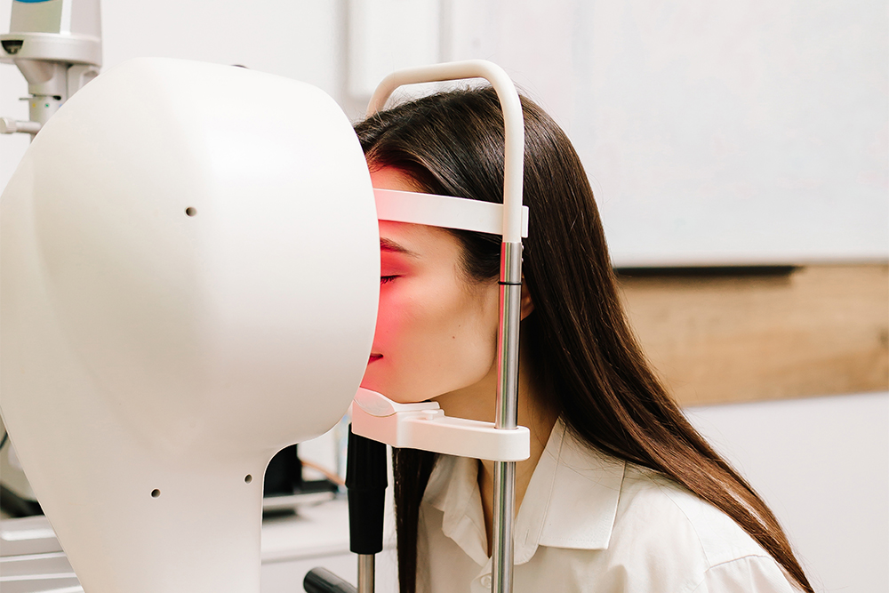
When it comes to diagnosing and managing complex vision conditions, precision matters. One of the most advanced tools is Medmont topography - a state-of-the-art technology that provides highly detailed maps of the cornea. Whether you are being evaluated for specialty contact lenses, dry eye treatment, or refractive surgery, Medmont topography plays a critical role in delivering accurate, customized care.
What Is Medmont Topography?
Medmont topography refers to corneal imaging performed with the Medmont E300 Corneal Topographer, a diagnostic device designed to capture detailed curvature measurements of the cornea.
Using advanced Placido disc technology, the system projects illuminated rings onto the cornea and analyzes their reflections. From this data, it generates a color-coded map that reveals the shape, elevation, and curvature of the cornea with exceptional accuracy.
Why Is Corneal Topography Important?
The cornea is responsible for approximately two-thirds of the eye’s focusing power. Even subtle irregularities can significantly affect vision. Medmont topography allows your eye doctor to detect and monitor:
Keratoconus and other corneal ectasias
Corneal irregularities or scarring
Post-surgical corneal changes
Astigmatism patterns
Contact lens fitting complications
Early signs of corneal distortion
Because it provides highly detailed imaging, Medmont topography often identifies changes long before they become noticeable in a routine vision exam.
How Medmont Topography Improves Contact Lens Fitting
For patients who require specialty contact lenses - such as scleral lenses, hybrid lenses, or lenses for keratoconus - precise corneal mapping is essential.
The Medmont system offers:
High-resolution curvature data
Wide corneal coverage
Repeatable, reliable measurements
Advanced software analysis
This allows your doctor to design and fit custom lenses with greater accuracy, improving comfort, stability, and visual clarity while reducing trial-and-error fittings.

Optical Coherence Tomography is a non-invasive imaging test that may be performed as a standard part of your regular, comprehensive exams, or you may be able to request this test as an addition to your usual exam.
Optical Coherence Tomography uses light waves to take cross-section images of your retina, which is the area of light-sensitive cells at the back of your eye that is responsible for receiving light and transmitting it into messages that are sent up to the brain. The technology behind OCT enables your eye doctor to see each of the different layers that make up the retina. By being able to see these and measure them, they can obtain a much clearer picture of the overall health and condition of your eyes.
Why are Optical Coherence Tomography scans important?
When you choose to have an OCT scan at fairly regular intervals, such as during your normal comprehensive eye exams, your eye doctor can compare newer results to previous ones. This helps them to build up a picture of the health of your eyes, and spot any changes which may be concerning, early, before they cause symptoms or have a permanent effect on your vision.
Anyone can have an OCT scan, but they are particularly recommended for patients over the age of 25 who are concerned about the health of their eyes, or who are at risk of or already have diabetes, glaucoma or a family history of eye disease. This is because they can be used to spot the early signs of a range of eye diseases, including glaucoma, diabetic retinopathy, macular degeneration, disorders of the optic nerve and more – even before you realize that you are affected.

Dry eye isn’t always just “not enough tears.” Often, the surface of the eye is irritated and inflamed, so tears don’t stay smooth or stable. For some people with more severe symptoms, amniotic membrane therapy can help by acting like a protective bandage that calms the surface and supports healing.
What Are Amniotic Membranes?
Amniotic membranes are derived from the innermost layer of the placenta and are carefully processed for medical use. Amniotic membranes are used to support regeneration of the corneal surface.
They contain growth factors and biologically active components that:
Reduce inflammation
Promote tissue healing
Minimize scarring
Protect the surface of the eye
Because they are immune-privileged and thoroughly screened, they are considered safe and well.
How Do Amniotic Membranes Help Dry Eye?
In moderate to severe dry eye disease, chronic inflammation can damage the corneal surface. This damage can lead to persistent discomfort, fluctuating vision, and delayed healing.
Amniotic membranes work by acting as a biological bandage. Once placed on the eye, they:
Create a protective barrier over the cornea
Reduce surface inflammation
Encourage regeneration of healthy cells
Restore smoother corneal tissue
By addressing the underlying inflammation and surface damage, amniotic membranes do more than simply mask symptoms - they promote healing at the source.



Contact Info
Office Address
Fax Number
(760) 727-3044
(760) 727-3044
Office Hours
- Monday 9:00am - 6:00pm
- Tuesday 9:00am - 6:00pm
- Wednesday 7:00am - 5:00pm
- Thursday 9:00am - 6:00pm
- Friday 9:00am - 2:00pm
- Saturday Closed
- Sunday Closed
© 2026 Shadowridge Family Vision Center. All rights Reserved. Accessibility Statement - Privacy Policy - Sitemap
Powered by:
